UB是一所一流的、研究型的公立大学,致力于学术卓越。我们的思维、研究、创造性活动和人们对世界产生了积极的影响。|了解有关UB的更多信息

发生在#ubuffalo.
在YouTube上
-
没有一个领域可以解决气候变化。了解UB如何教学学生,使联系会产生差异。
即将举行的活动
加载活动......
推特
Instagram.
自豪点
-
第1号
被《美国新闻与世界报道》认定为纽约州顶尖公立大学,全国排名第34位。
-
前1%
在世界大学排名中心全球19788所大学中排名第293位。
-
前40名
在《华尔街日报》和《美国新闻与世界报道》的大学排名中,它是美国第31位公立大学,在《美国新闻与世界报道》中,它是第34位。
-
主要选择
东北地区的第2位公立大学,为最广泛的计划(450+)。
-
世界级教师
诺贝尔奖获奖者,普利策奖,Macarthur基金会“Genius奖”和(这么多)其他人。
-
个人关注
14:1的师生比例——在纽约州立大学所有授予博士学位的大学中排名第一。
-
最大的图书馆
UB的图书馆和特殊收藏中有超过400万卷 - 任何Suny机构的大多数。
-
经验丰富
所有学生从第一年都可以参与研究,服务或经验学习。
-
荣誉学院
全国首批荣誉课程之一,也是纽约州立大学皇冠上的一颗宝石。
-
在国外学习x 1,000
任何主要的学生都可以通过七大洲的1,000多种选项出国留学。
-
新的音乐领袖
6月份在布法罗,世界知名的新音乐节和会议。
-
启动文化
学生企业家在众多年度比赛中竞争启动资金和服务。
-
精英中的精英
作为AAU成员,在北美北美大学中排名,从事最高水平的研究。
-
东北部的第四名
由WSJ /大学排名认可为东北部第四届最佳公立大学。
-
“最佳价值”
由Kiplinger和美国新闻和世界报告称为高等教育最佳价值之一。
-
完成4.
通过遵循我们的指导方针 - 或剩余的学费确保学生毕业。
-
DI田径
16个在中美洲联盟竞争(并获胜)的NCAA一级运动项目。
-
准备成功
90%的布法罗大学本科毕业生在研究生院工作。
-
梦想的工作
谷歌,美国宇航局,辉瑞,U.N.,CNN,史密森尼,花旗,Spotify,HBO等,UB毕业的土地工作。
-
晴天
东北部任何主要城市最阳光最干燥的夏天。
-
我们着名的家庭
世界领导人,宇航员,NFL传说,庆祝的记者,美国诗人洛杉矶和清单。
-
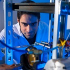
研究排名前35位
全国领先的公共研究大学之一,每年花费400 00 +百万美元。
-
NYS中最好的研发
纽约州顶级公立研究与开发。
-
大型奖学金
283岁的奖学金在过去35年中获胜,包括杜鲁门,Goldwater和Marshall。
-
卓越的价值
具有学费的顶级教育,这是一个可比私立学校成本的一小部分。
-
数百万人艾滋病
每年在经济援助和奖学金中超过3.6亿美元。
-
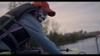
免费皮划艇
一个60英亩的湖泊,坐在校园中间,配有免费皮划艇,独木舟和桨板。
-
oozefest.
美国最大的大学泥浆排球比赛之一。
-
好吃
Suny的1号餐饮服务和一致的餐饮奖奖获得者。
-
自然浸没
65英亩的生活伍兹在校园里。
-
做出改变
高影响力的经验机会,都在巴菲罗和世界各地。
-
全球美国
该国最国际大学之一,为学生提供真正的全球经验。
-
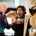
多元化的历史
毕业于18世纪后期的医学院的第一名女性和黑人学生。
-
妇女负责
一半的UB院长是女性,包括法律院长和艺术与科学的院长。
-
民权冠军
抗议种族主义,我们庆祝的1958年足球队队拒绝了UB的第一个碗游戏邀请。
-
绿色领袖
EPA的绿色电力伙伴关系名单第4名高等学校。
-
由太阳提供动力
3,200面板UB太阳能钢铁生产足够的能量来推动数百名学生公寓。
-
气候活动人士
根据“较高的教育冲击排名,气候行动的第1名气候行动和2号清洁能源。
-
野生动物欢迎
通过认证的野生动物栖息地认识到校园众多物种的野生动物栖息地计划。
-
校友到处都是
所有50个州和150个国家的273,000多人有用的UB校友。
-
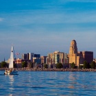
一个城市上升
在不断增长的荣辱程度中:一个“十大美国城市”(福布斯)和“最佳旅行”(孤独的星球)
-
经济实惠的生活
布法罗名为“25个最实惠的地方”之一(美国新闻和世界报告)。
-
达到的海滩
驱车30分钟即可到达两大湖沿岸的众多海滩。
-
庆祝活动加罗尔
每年100多个地区节日庆祝从爵士乐到侏儒的一切。
-
最好的邻居
正如纽约时报作者杰达元在访问水牛后说:“我发现了一种似乎在化妆中根深蒂固的慷慨。”
-
大脑博物馆
美国唯一一个专门研究人脑的博物馆。
-
历史生活在这里
UB的持股包括火箭带,早期纸张,原始的“孤独的游侠”无线电剧本。
-
LGBTQ + Pioneer.
UB的“Lesbianism 101”是美国在美国教授的第一课程